

Atoms are the basic building blocks of materials. The three-dimensional (3D) arrangement of atoms determines the physical properties of matter. To understand material properties and functionality at the most fundamental level, one must know the 3D positions of atoms with high precision. However, perfect crystals are rare in nature. The properties of many materials depend on vacancies, defects, surface reconstructions, grain boundaries, stacking faults, dislocations and disorder. Those atomic arrangements are not accessible to crystallography. More importantly, non-crystalline structures can evolve into different structures under external stimuli, but their atomic-scale dynamics are currently poorly understood. Our research interests entail using and developing advanced electron microscopy, particularly atomic resolution electron tomography, to solve traditionally intractable problems in physics, chemistry and materials science.
Interface is the device.
1 Metal-oxide interface
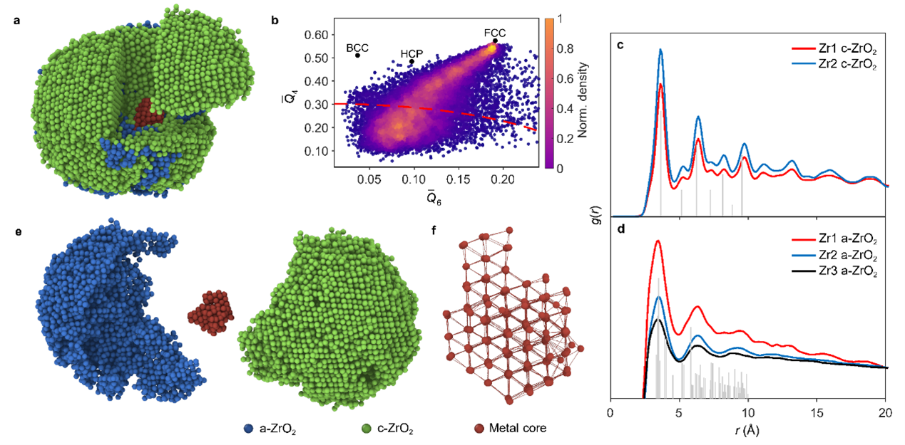
Metal-oxide interfaces with poor coherency have specific properties comparing to bulk materials and offer broad applications in heterogeneous catalysis, battery, and electronics. However, current understanding of the three-dimensional (3D) atomic metal-oxide interfaces remains limited because of their inherent structural complexity and the limitations of conventional two-dimensional imaging techniques. Recently, we determine the 3D atomic structure of metal-oxide interfaces in zirconium-zirconia nanoparticles using atomic-resolution electron tomography (AET) [1].
Using Zr-ZrO2 nanoparticles as a model system, we determined the 3D atomic structure of the metal-oxide interface using AET. We quantitatively measured the atomic packing density and the degree of oxidation from our experimental model of the metal-oxide interface. The degree of oxidation from metal to oxide increases gradually, resulting in a diffuse interface between FCC Zr core and ZrO2. The Zr metal connects with its oxide via {111} planes; and the semicoherent interface has severe distortion, including bending and twisting. The significant stress in the interface is relieved through low coordination and defects. Numbers of defects, including vacancies and nano-pores, together leverage the mass transportation during oxidation.
We anticipate that our findings will fulfill the dearth of 3D atomic structure of metal-oxide interface and advance the study of fundamental problems of metal-oxide interfaces such as oxidation kinetics, diffusion, and defect evolution in variety of materials.

2 Metal-metal interface
3D structural analysis is essential to understand the structure and property relationships of materials. A disruption in periodicity strongly affects material properties and functionality. Atomic Electron Tomography (AET) can determine the 3D structure of crystal defects and disordered materials such as grain boundaries, stacking faults, the core structure of edge and screw dislocations, point defects and chemical order/disorder at atomic resolution. Deciphering the three-dimensional atomic structure of solid-solid interfaces in core-shell nanomaterials is the key to understand their catalytical, optical and electronic properties.
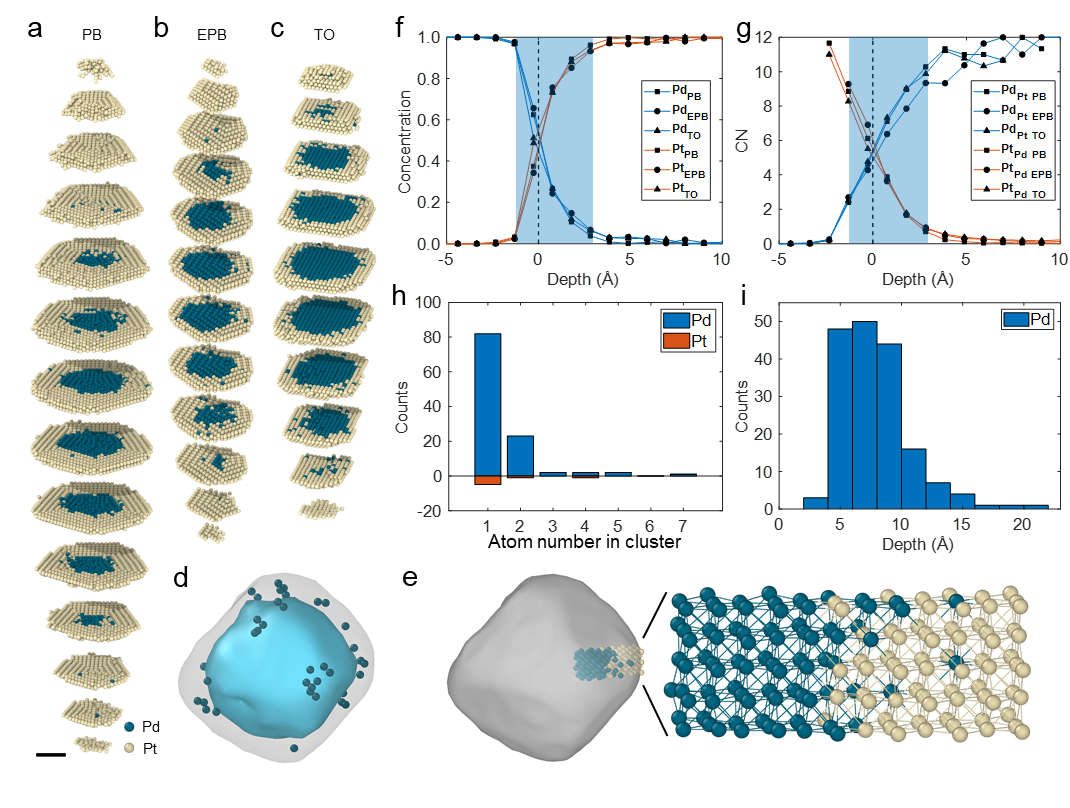
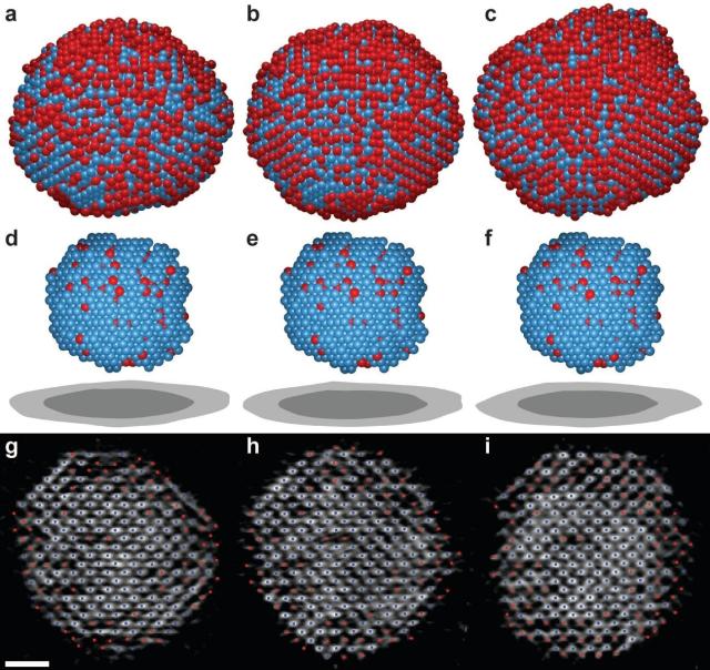
Recently, we probe the three-dimensional atomic structures of palladium-platinum core-shell nanoparticles at the single-atom level using atomic resolution electron tomography [2]. We quantify the rich structural variety of core-shell nanoparticles with heteroepitaxy in 3D at atomic resolution. Instead of forming an atomically-sharp boundary, the core-shell interface is found to be atomically diffuse with an average thickness of 4.2 Å, irrespective of the particle’s morphology or crystallographic texture.
The high concentration of Pd in the diffusive interface is highly related to the free Pd atoms dissolved from the Pd seeds, which is confirmed by atomic images of Pd and Pt single atoms and sub-nanometer clusters using cryogenic electron microscopy. These results advance our understanding of core-shell structures at the fundamental level, providing potential strategies into precise nanomaterial manipulation and chemical property regulation.
[1]. Zhang, Y. et al., Three-dimensional atomic insights into the metal-oxide interface in Zr-ZrO2 nanoparticles, Nat. Commun. 15, 7624, 2024. [link]
[2]. Li, Z. et al., Probing the atomically diffuse interfaces in Pd@Pt core-shell nanoparticles in three dimensions, Nat. Commun. 14, 2934, 2023. [link]
While the 3D static atomic structure of materials is important to understand their functionality, there exists significant interest to reveal the structure and dynamics of materials at 4D atomic resolution to study processes such as nucleation and growth. We developed 4D (the three dimensions of space plus the fourth dimension of time) atomic electron tomography to directly image the dynamics of structural changes at the atomic scale.
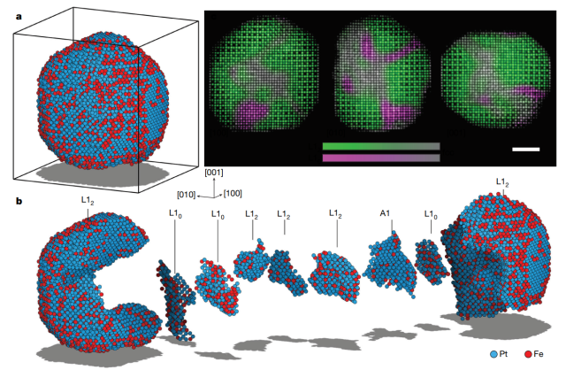
Nucleation is the process by which tiny clusters of atoms or molecules (called “nuclei”) begin to coalesce. Nucleation plays a critical role in phenomena as diverse as the formation of clouds and the onset of neurodegenerative disease. Using FePt nanoparticles as a model system, Zhou et al. have recently studied the dynamics of early stage nuclei in an ex-situ AET experiment [3]. The initiation of nucleation on the surface of the nanoparticles is observed and the structure and dynamics of the same nuclei undergoing growth, fluctuation, dissolution, merging and/or division is captured. These results not only show a never-before-seen view of nucleation but also indicate that a theory beyond classical nucleation theory is needed to describe early-stage nucleation at the atomic scale. This experiment adds a new dimension (time) to AET (i.e. 4D AET), capturing atomic motion in materials in four dimensions, which is currently not accessible by any other experimental methods.
4D AET will potentially serve as a powerful tool in studying many fundamental problems such as phase transitions, atomic diffusion, grain boundary dynamics, interface motion, defect dynamics and surface reconstruction.
[3]. J. Zhou et al., Observing crystal nucleation in four dimensions using atomic electron tomography, Nature, 570, 500-503, 2019. [link]
The deepest and most interesting unsolved problem in solid state theory is probably the theory of the nature of glass and the glass transition. This could be the next breakthrough in the coming decade.
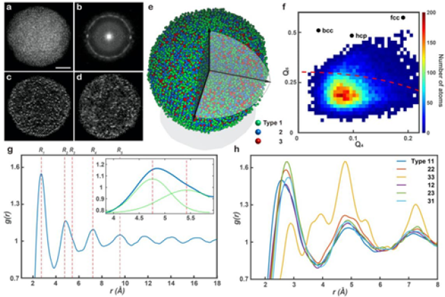
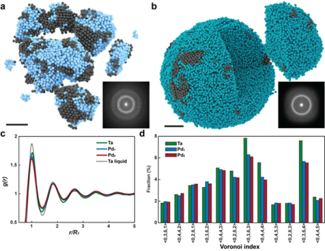
Amorphous materials such as glass are ubiquitous in our daily life and have broad applications ranging from window glass and solar cells to telecommunications and transformer cores. However, due to the lack of long-range order, the 3D atomic structure of amorphous materials have thus far defied any direct experimental determination without model fitting. Recently, with a breakthrough of atomic resolution electron tomography experiment, we are able to quantitatively characterize amorphous solids and disorder structures at 3D atomic resolution.
Using a multi-component glass-forming alloy nanoparticle in which a large portion of atoms have disorder structures as proof of principle , we characterize the short-range order and medium-range order of the 3D atomic arrangement [4]. Although the 3D atomic packing of the SRO is geometrically disordered, some SRO connect with each other to form crystal-like networks and give rise to MRO. These observations confirm that the 3D atomic structure in some parts of the sample is consistent with the efficient cluster packing model.
The ability to determine the 3D atomic structure of amorphous materials [4,5] could open a door to study the holy grail problem in condense matter physics -- the structure of glass and the nature of glass transition.
[4]. Yang, Y. et al., Determining the three-dimensional atomic structure of an amorphous solid, Nature 592, 60-64, 2021. [link]
[5]. Yuan, Y. et al., Three-dimensional atomic packing in amorphous solids with liquid-like structure, Nat. Mater. 21, 95-102, 2022. [link]